
Cancer is a scary word. At its core, it’s a coup: almost any cell in the body can mutate or become damaged, start to grow uncontrollably, and metastasize (spread and take root) to other organs if not stopped in time.
It’s this metastasis that accounts for up to 90% of cancer-related deaths.
Dr. Jing-Fei Dong at Bloodworks Research Institute is conducting research on how cancer metastasizes and how to recognize mechanisms that allow cancer cells to invade healthy organs. While solid tumors do not originate in the blood, they spread to new parts of the body through blood circulation.
“Blood is the only organ that is everywhere, and it is fluid,” Dr. Dong said. “It goes everywhere, but it doesn’t stop there. It is constantly circulating. It carries things to and from places. It’s function, and it’s disfunction, is probably the pathology behind many if not all diseases.”
Identifying these factors may help develop new treatments to block the spread of cancer.
Blood vessels in cancer tissue are weak and prone to “leaks” and breakage that allow cancer cells to release into blood circulation.
However, just because something can get out of a faulty vessel doesn’t mean it should be able to get in to a healthy one.
Your heart, blood vessels, and lymphatic system are lined by a layer of cells called endothelial cells that form a selectively permeable wall called the endothelium. The endothelium helps blood flow and is the critical barrier between your bloodstream and the other tissues in your body. It lets nutrients in and releases waste products (cellular transport) and keeps out the harmful stuff (like cancer cells).
In theory, the endothelium should stop cancer from spreading to new organs as part of the body’s defense system and immune response.
But this isn’t always the case.
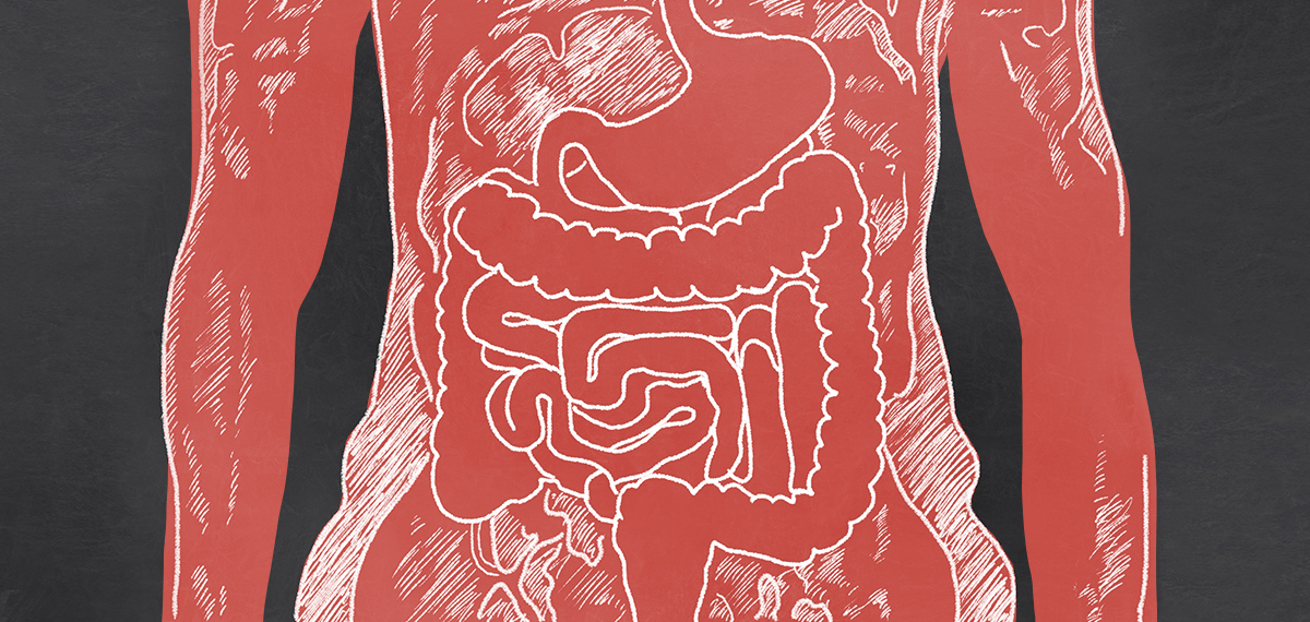
Different types of cancers often have different primary targets for metastasis. Stomach cancer tends to spread to the lungs and liver.
Most people with early stage stomach cancer have a relatively good prognosis, but once it has spread it becomes harder to treat. The five-year survival rate in patients whose cancer has metastacized from the stomach to the liver is only 6%.
How do cancer cells break otherwise healthy vascular barriers to form new cancer masses? And how does stomach cancer spread specifically to the lungs and liver?
The answers are in the blood.
The Dong Lab collaborated on an exciting study that looked at how cancer cell-derived extracellular vesicles (CEVs)—tiny protrusions produced by the membranes of cancerous cells that play a role in regulating cellular transport—regulate cancer metastasis by analyzing samples from gastric (stomach) and cervical cancer patients, performing lab-based experiments, and studying living organism models.
The study reveals the role of CEVs in promoting the metastasis of gastric cancer to the lungs.
This research shows that metastasis is linked to plasma levels of CEVs in patients with gastric cancer. Subjects with pulmonary (lung) metastasis showed higher levels of CEVs circulating in their plasma than those without. Patients with gastric cancer had significantly higher levels of CEVs in their plasma than the patients with cervical cancer.
It also shows how gastric cancer specifically spreads to the lungs. CEVs disrupt the endothelial barrier to promote cancer metastasis. Healthy endothelial cells endocytose (surround and eat) CEVs, resulting in cytoskeletal rearrangement (damage to the healthy cell’s exterior wall), reduced function of cadherin and CD31 (proteins that regulate osmosis and other passage across cell membranes). Cadherin helps our heart and lungs respond to protective signals, so a reduction of cadherin levels has significant effects on these particular organs—hence, why the lungs are so impacted.
CEVs also cause the formation of large gaps between cells, which allow cancer cells to move across the endothelium.
Finally, the dynamin (a protein essential for cell membrane function) inhibitor Dynasore prevents CEV-induced changes of endothelial cells by blocking CEV endocytosis. Lactadherin, which was originally discovered in mold, removes CEVs from the circulation to preserve the integrity of vascular wall and thus prevent cancer metastasis.
Understanding how disease spreads can often be the key to stopping it. Because metastasis is associated with high gastric cancer mortality, the medical possibilities these findings reveal are huge, as is the potential to save lives.
These results also suggest that the medically accepted relationship between lactadherin and cancer may be incomplete: previous studies have negatively linked lactadherin levels with tumor growth and mortality in breast cancer patients. One difference may be that previous studies evaluated the effects of cancer cell-derived lactadherin at the primary cancer site rather than how circulating lactadherin impacts tumor metastasis. It could also suggest that cancer patients with low levels of scavenging molecules, like lactadherin, might have a higher risk of metastasis.
The questions both could be an opportunity for further research—and potentially future pharmaceutical breakthroughs.
The Dong lab has concluded a second study that builds on this. This research shows that CEV-mediated metastasis uses von Willebrand factor (VWF), a platelet-clotting protein on which Bloodworks is the leading expert.
Blood truly touches every disease. By examining the critical role blood plays in every area of the body, we’re able to better understand the overall picture of human health.
Your support makes a difference in funding breakthroughs like this one.


Tell Us What You Think!